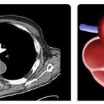
Pulmonary stenosis is a narrowing of the outlet from the right ventricle or pulmonary artery valve and is classified according to its severity.
What is pulmonary stenosis?
Pulmonary stenosis is a narrowing of the outflow path between the right ventricle and the pulmonary artery. The pulmonary valve is located between the pulmonary artery and the right ventricle. Through them, the oxygen-poor blood reaches the lungs. The flap is therefore a valve that is responsible for controlling blood flow. It is a congenital heart defect that either occurs in isolation or can also be part of a complex heart defect. See topbbacolleges for Definitions of Bone Marrow Failure.
There are three different types of pulmonary stenosis:
- subvalvular pulmonary stenosis: narrowing of the outlet from the right ventricle due to excess tissue
- valvular pulmonary stenosis: affects the valve itself, where the valve pouches are partially fused or thickened and the valve opening is not complete.
- Supravalvular pulmonary stenosis: narrowing above the valve and narrowing of the pulmonary artery
The most common type is valvular pulmonary stenosis, which affects more than 90 percent of cases.
Causes
In many cases, pulmonary stenosis is a congenital heart defect, the causes of which are not known. Under certain circumstances, however, a genetic predisposition can be held responsible. It is also possible that the pulmonary valve is not fully developed during pregnancy. In addition, pulmonary stenosis can also occur in addition to a congenital heart defect or in the course of rheumatic fever or cancerous tumors in the digestive tract.
Symptoms, Ailments & Signs
The symptoms of pulmonary stenosis vary greatly and depend on the severity of the narrowing. If the constriction is only very slight, there are usually no symptoms. In severe cases, shortness of breath (dyspnoea) occurs, which is particularly noticeable when the heart is under stress. In addition, those affected suffer from peripheral cyanosis, which means that the patients are not supplied with sufficient oxygen.
The heart is unable to pump enough deoxygenated blood to the lungs. As a result, the red blood cells, which are responsible for transporting oxygen and exchanging it for carbon dioxide, do not receive any new oxygen. Thus, it is not possible for them to release the carbon dioxide. Peripheral cyanosis can be detected with the help of a blood test, in which case the level of carbon dioxide in the red blood cells is greatly increased.
For the heart, the constant attempt to pump blood through the heart valve means an extremely great effort. As a result, the blood presses on the heart muscle, which grows because it has to adapt to the pressure conditions. If the narrowing of the heart valve is very severe, heart failure can also occur. Other possible symptoms include fatigue, a bulging abdomen, fainting, and a bluish discoloration of the skin.
Diagnosis & course of disease
Pulmonary stenosis can be diagnosed in different ways. First, the doctor listens to the patient with a stethoscope. As a result, he hears the heart sounds, whereby a so-called split second heart sound can be heard in the case of pulmonary stenosis, which is due to the narrowing. A murmur called the “systolic” can also be heard as blood rushes out of the ventricle.
An ECG is also very often carried out, whereby changes can be detected in the event of a severe narrowing. Another examination method is the echocardiogram. This is an ultrasound scan that allows the doctor to visualize the structure of the heart. The heart or the heart valves can be viewed on a monitor and the direction of flow of the blood can be determined with the help of a color Doppler.
An enlarged right heart can also be seen on an X-ray. The pulmonary vessels, on the other hand, are only shown very faintly, which is a sign that only little blood is being transported through the narrowed heart valve into the lungs. A so-called invasive method is a right heart catheter, which can provide very precise information about a possible heart defect. With the help of a catheter, it is possible to assess the severity of the narrowing. To do this, the doctor inserts a catheter into a vessel on the thigh and then advances it to the heart, where the tip of the catheter can measure the pressure conditions in the pulmonary artery or the ventricles of the heart.
Complications
In most cases, those affected by pulmonary stenosis suffer from heart problems or breathing difficulties. The resilience of those affected by the disease also drops significantly and the patients become permanently tired and exhausted. The internal organs are also supplied with less oxygen due to the pulmonary stenosis and can be damaged as a result.
In the worst case, it can also lead to carbon dioxide poisoning of those affected. Since the heart also has to pump an increased amount of blood, heart failure or other heart problems can occur. In the worst case, the person concerned dies of heart failure. As a rule, without treatment, the patient’s life expectancy is significantly reduced. This disease can be treated by surgery.
There are no particular complications. However, the affected person can no longer perform strenuous activities or sports. Furthermore, the patient is also dependent on medication to prevent further complaints. With successful treatment of pulmonary stenosis, life expectancy is not affected in most cases. A healthy lifestyle can also have a very positive effect on this disease.
When should you go to the doctor?
Pulmonary stenosis must always be treated by a doctor. In the worst case, the person affected can die, so that early diagnosis and treatment always have a very positive effect on the further course of the disease. Pulmonary stenosis usually manifests itself as shortness of breath. Shortness of breath can occur, especially during strenuous activities or sporting activities, and the affected person can also lose consciousness completely. Cyanosis can also be indicative of pulmonary stenosis and should be investigated if it occurs over a long period of time and reduces the patient’s quality of life. Furthermore, permanent tiredness or a strongly protruding abdomen also indicate the disease and must be examined by a doctor.
First and foremost, the disease can be examined by a general practitioner or by a cardiologist. However, if an emergency or loss of consciousness occurs, an ambulance should be called or the hospital should be visited.
Treatment & Therapy
A frequently chosen method for the therapy of pulmonary stenoses is an expansion of the constricted heart valve with the help of a balloon. The balloon is placed at the same level as the pulmonary stenosis using a cardiac catheter and then inflated. As a result, the altered heart muscle can be reformed. In the case of very severe stenoses, however, an operation may also be necessary.
During this operation, the pulmonary valve is reconstructed or a heart valve is inserted. Newborns suffering from severe pulmonary stenosis require intensive medical care. In addition, the doctor may prescribe medications that improve blood flow. These include, for example, medication for cardiac arrhythmias, water pills to enable increased water excretion, blood thinners and prostaglandins that improve blood circulation.
Prevention
Since pulmonary stenosis is a very common congenital heart defect, it cannot be prevented. However, those affected should lead a heart-friendly and healthy lifestyle and avoid cigarettes. A healthy diet and regular exercise are also important.
Aftercare
The different degrees of severity and the causes of pulmonary stenosis lead to different forms of therapy. The spectrum of possible treatment ranges from a change in diet to balloon dilatation, insertion of a stent and surgical replacement of the pulmonary valve in the right ventricle. The need for follow-up treatments and follow-up examinations is correspondingly differentiated.
Starting with a milder form of pulmonary stenosis, the primary need for follow-up examinations arises. This is used to determine whether the severity of the stenosis has decreased over the long term or whether the disease is progressing so that further treatment or surgery is indicated. The most important diagnostic devices for follow-up examinations are the stethoscope, EKG and the Doppler ultrasound machine.
Regular follow-up examinations are also recommended after balloon dilatation or endoprosthetic replacement of the pulmonary valve. Doppler sonography is of particular importance as a follow-up examination. It can thus be tracked whether a thickening of the heart wall of the right ventricle (hypertrophication) is regressing, which can be taken as an indication that the intended therapeutic purpose has been achieved.
Further follow-up examinations are recommended from time to time, since renewed narrowing of the pulmonary valve often occurs without symptoms at first. There is a risk that the renewed narrowing of the pulmonary circulation will not be noticed until very late, which can make subsequent therapy more difficult.


