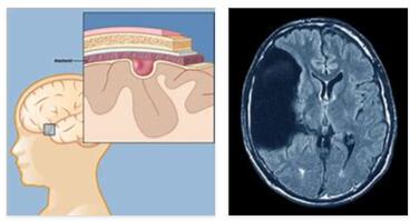
In the case of the arachnoid cyst, the medical practitioner understands an accumulation of liquor that is surrounded by the arachnoid (spider web-like meninges). The human brain has three layers of meninges, the middle layer of which consists of thin and white collagen fibers.
What is an arachnoid cyst?
According to ablogtophone, Arachnoid cysts are often found as congenital malformations in the arachnoid (spider web-like meninges) that arise in the third trimester during early childhood development.
The term arachnoid cyst describes a cavity in the spider web skin (arachnoidea) that is filled with nerve water (liquor). This brain-water-filled, factual protuberance of the meninges can be congenital or result from external effects of injuries, operations or diseases. Most of the time they go undetected, but they can also increase in size and cause discomfort.
The cysts are differentiated between intracranial and spinal meningeal. An intracranial arachnoid cyst is located within the skull or inside the skull. The spinal meningeal arachnoid cysts lie within the spine (intraspinal) but outside the spinal cord (extramedullary). They contain cerebrospinal fluid ( cerebrospinal fluid ).
Causes
Arachnoid cysts are often found as congenital malformations in the arachnoid (spider web-like meninges) that arise in the third trimester during early childhood development. Reasons for the formation of cysts can be medication, drugs or the effects of radiation exposure. Arachnoid cysts are also associated with the effects of meningitis .
Due to malformations, a blister-like structure develops in the middle meninges in which cerebrospinal fluid (cerebrospinal fluid) collects. As the water increases, an arachnoid cyst forms, which can be found either spinally or intracranially. Arachnoid cysts often show near the Sylvian fissure.
Other causes include inflammation of the brain (meningitis) and the nerve cell fibers of the spinal cord, circulation problems between the right and left hemispheres (corpus callosum) or the inherited autosomal dominant disease of the connective tissue ( Marfan syndrome ).
Symptoms, ailments & signs
Arachnoid cyst symptoms are usually rare. However, a large number of the cysts are discovered by chance as a result of an MRI or CT examination. As soon as an arachnoid cyst increases in size and thereby displaces surrounding tissue and organs, symptoms are indicated. Depending on the localization, it manifests itself in very differentiated symptoms.
Cystic enlargements can occur in various places intracranially (within the skull), most commonly in the area of the temporal brain. Sometimes they also occur in the area of the Sella region behind the cerebellum. Intracranial arachnoid cysts can sometimes cause symptoms such as headaches , increased intracranial pressure with nausea and vomiting .
They can also trigger increased fatigue , epileptic seizures , visual disturbances , speech disorders or symptoms of developmental delay as well as hormonal disturbances in the form of premature puberty development. Sometimes patients also stand out due to personality changes .
Spinal arachnoid cysts can negatively affect the spinal cord and the circulation of liquor by permanently exerting pressure on the nerve roots. Depending on the location, the pressure on the spinal cord or nerve roots causes radiating pain in extremities such as arms and legs, unsteady gait , and sensitivity disorders . Symptoms of paralysis or disorders of the bladder and bowel function.
Sometimes external injuries are also decisive for the development of a cyst. In particular, minor injuries can lead to bleeding. However, there are often no symptoms whatsoever, so that the arachnoid cyst is discovered more by chance.
Diagnosis & course
If an arachnoid cyst is suspected, two important methods are used. Magnetic resonance tomography is often recommended due to the exact representation of the soft tissue . In this process, a three-dimensional model of the vessels is created using magnetic field technology, which specifically detects enlargements in the brain or spinal cord.
The cyst shows up as a fluid-filled space. In rare cases, the condition between the cyst and the outer cerebral water spaces is also examined by enriching the cerebral fluid with a contrast agent . The content of the cyst is shown in the MRI with the same intensity as that of the cerebrospinal fluid (liquor cerebrospinales) and the contrast agent clearly shows the cyst wall. Adjacent tissue structures are included to differentiate the diagnosis.
As an alternative method, the ultrasound examination is the focus, especially for infants and children. The reflecting sound waves create images of structures within the body. Diagnostic sonography of arachnoid cysts takes only a few minutes.
Once the presence of cysts is diagnosed, the doctor will take further steps. In a subsequent neurological examination, the functions of the brain and spinal cord are checked in order to be able to rule out possible, as yet unnoticed impairments. This is followed by reviews of the patient’s reflexes, sensitivity, and motor functions. A blood sample with determination of the inflammation parameters also provides evidence of an arachnoid cyst.
Complications
In most cases, the arachnoid cyst will only experience discomfort and symptoms when the cyst grows. For this reason, only a delayed or accidental diagnosis is usually possible. In most cases, the enlargement of the cyst leads to severe headaches due to the increased pressure in the brain.
The patients also suffer from vomiting and nausea and are extremely limited in their everyday life. The severe headache can lead to difficulty concentrating or sleeping. Epileptic seizures also occur, which are associated with fatigue. As a rule, it is not possible to compensate for this tiredness with sleep.
Visual disturbances can also occur suddenly. In children, development and intelligence disorders occur due to the arachnoid cyst. In some cases, the personality or basic attitude of a patient can change significantly, which can have a negative impact on social contacts. Furthermore, paralysis occurs in various parts of the body.
These can lead to restricted mobility or to gait disorders. Often the patients also suffer from word-finding disorders and language disorders. The complications can persist even after the cyst is removed if it is done late. With an early removal, consequential damage can be avoided.
When should you go to the doctor?
An arachnoid cyst does not necessarily require treatment. However, should symptoms arise, a doctor must diagnose the cyst and remove it if necessary. Typical symptoms such as headache, impaired vision and language, nausea and vomiting, fatigue and epileptic seizures suggest a serious cause.
Although it is rarely an arachnoid cyst, there is almost always an illness that must be clarified and treated by a doctor. It is therefore advisable to see a doctor if you have unusual symptoms that persist for more than a few days.
A diagnosed arachnoid cyst should be observed. If you suddenly experience hormonal imbalances, symptoms of a developmental disorder, or the above symptoms, the cyst may have enlarged. At this point at the latest, the growth should be surgically removed.
Other warning signs that require immediate medical clarification are sensitivity disorders, symptoms of paralysis and insecurities in the hallway. Also sudden onset of pain in the limbs and disorders of the bladder and bowel function. If these complaints arise, the following applies: go to the family doctor or neurologist and have the cause determined.
Treatment & Therapy
Treatment and therapy first require a causal clarification. To do this, it is necessary to carefully examine the current general condition of the patient and the risk factors for the corresponding therapy. Arachnoid cysts should only be treated surgically if symptoms arise.
Cysts discovered by chance require regular check-ups using imaging techniques. However, as soon as the cyst causes a displacement that affects the blood circulation or the circulation of the cerebrospinal fluid, surgical treatment is necessary. Various methods are available for this.
The endoscopic cyst windowing is a minimally invasive and at the same time gentle method. The cyst wall is opened endoscopically in order to establish a broad connection (marsupialization) to the natural cerebral water spaces of the brain base (cisterns) or to the brain chambers (cysto-ventriculo-stomy). to accomplish. If the cyst wall is firm, the microsurgical technique is required.
In rare cases, a cystoperitoneal shunt is applied . This is a catheter that is inserted into the cyst. This directs the fluid under the skin using a pressure valve up to the abdomen, where the draining cerebral fluid is absorbed. This method achieves an enormous relief of the brain water drainage disorder.
Outlook & forecast
The prognosis for an arachnoid cyst is generally good. In the majority of cases, the cyst is removed immediately after the diagnosis is made without further impairment and the patient can be discharged as cured. The procedure is usually uncomplicated and possible with little effort. The subsequent wound healing takes several weeks. After that, the person affected is symptom-free.
This prospect of cure depends on the location of the cyst’s appearance, the size of the cyst, and the patient’s health. The older the person concerned is and the more previous illnesses there are, the less favorable the healing process. Nevertheless, the arachnoid cyst is usually completely and permanently removed here as well.
If the cyst is in an area that is difficult to access, the likelihood of a good prognosis decreases. Removal can be associated with serious complications and lifelong impairment. The chances of recovery are also reduced if the arachnoid cyst has already caused functional disorders in the body. These can often no longer be corrected after removal. Without treatment, the cyst threatens to grow further. This increases the risk of physical disturbances and irreparable damage. In severe cases, the patient is threatened with life-threatening conditions.
Prevention
Congenital arachnoid cysts cannot be prevented through prevention. If their existence is known, however, regular checks using CT or MRI should be carried out. Arachnoid cysts that are based on existing diseases, such as high blood pressure in diabetes mellitus , can be limited by primary treatment of the underlying disease.
This also includes a conscious change in lifestyle or, in the case of pronounced high blood pressure, the use of drug therapy. With a timely diagnosis and subsequent operation, the quality of life of those affected can be increased enormously.
Aftercare
In most cases, arachnoid cysts are harmless and therefore do not require any intensive treatment or follow-up care. If the cysts have already been diagnosed, a follow-up should be carried out by the neurologist once or twice a year. If there are any complaints, surgery in a specialist clinic may be necessary.
After this procedure, follow-up care is limited to examining the brain with an ultrasound scan and monitoring the medication. In addition, it must be ensured that the removal of the cysts did not lead to neurological complaints. The patient should first take it easy and watch out for any unusual symptoms.
Aftercare also includes keeping a complaint diary. In this way, it can be tracked whether cysts have formed again after months or years. If this is not the case, follow-up care can be discontinued. In the event of a relapse, therapy can be resumed if the new cysts cause health problems.
Normally, arachnoid cysts are uncomplicated and only require irregular follow-up checks. Patients should contact their doctor if they experience discomfort after removing a cyst or if side effects or interactions occur as part of drug treatment.
You can do that yourself
Before an arachnoid cyst can be treated, a comprehensive evaluation must be carried out by the doctor. The actual therapy can be supported, among other things, by strict personal hygiene and compliance with medical guidelines.
Since the cysts can appear in several parts of the body, the patient should check the armpits, back, genital area and other areas of the body that are difficult to see for abnormal skin changes on a daily basis. An arachnoid cyst usually occurs in the brain area, but serious skin conditions that affect the entire body can develop with other conditions.
For this reason, a careful examination must be carried out by a specialist in neurology . People who have meningitis should inform their doctor.
Further measures depend on the type and severity of the cyst. Small cysts can be surgically removed and usually promise a positive prognosis. Larger cysts, on the other hand, can cause neurological disorders that must be treated separately. The patient should contact a physical therapist early on and take preventive measures. A therapeutic counseling reduces the risk of side effects of the psyche considerably.
")
